Showing posts with label CLASS 11 Biology (NCERT Solution). Show all posts

CLASS 11 Biology CHAPTER 9 Biomolecules (NCERT Solution)

CLASS 11 Biology 
CHAPTER 
Biomolecules (NCERT Solution)
 
Question 1: What are macromolecules? Give examples.
Answer: Macromolecules are large complex molecules that occur in colloidal state in intercellular fluid. They are formed by the polymerization of low molecular weight micromolecules.
Polysaccharides, proteins, and nucleic acids are common examples of macromolecules.

Question 2: Illustrate a glycosidic, peptide and a phospho-diester bond.
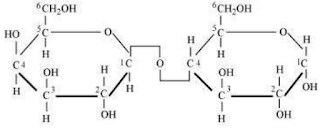
Answer: (a) Glycosidic bond is formed normally between carbon atoms, 1 and 4, of neighbouring monosaccharide units.


(b) Peptide bond is a covalent bond that joins the two amino acids by - NH - CO linkage.


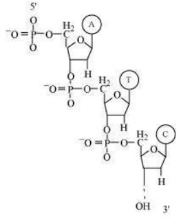
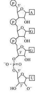
(c) Phosphodiester bond is a strong covalent bond between phosphate and two sugar groups. Such bonds form the sugar phosphate backbone of nucleic acids.


Question 3: What is meant by tertiary structure of proteins?
Answer: The helical polypeptide chain undergoes coiling and folding to form a complex three-dimensional shape referred to as tertiary structure of proteins. These coils and folds are arranged to hide the non-polar amino acid chains and to expose the polar side chains. The tertiary structure is held together by the weak bonds formed between various parts of the polypeptide chain.

Question 4: Find and write down structures of 10 interesting small molecular weight biomolecules .Find if there is any industry which manufactures the compounds by isolation. Find out who are the buyers.
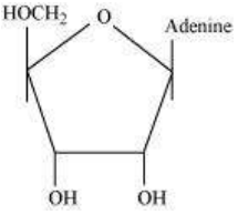
Answer: (a) 1. Adenosine


2. Thymidine


3. Sucrose


4. Maltose


5. Lactose
6. Ribose


7. DNA


8. RNA


9. Glycerol


10. Insulin


(b) Compound                    Manufacturer                      Buyer
1.  Starch products               Kosha Impex (P) Ltd.           Research laboratories, educational institutes,                                                                                               and other industries, which use biomolecules                                                                                               as a precursor for making other products.
2. Liquid glucose                 Marudhar apparels
3. Various enzymes              Map (India) Ltd
such as amylase, protease,
cellulase                                 

Question 5: Proteins have primary structure. If you are given a method to know which amino acid is at either of the two termini (ends) of a protein, can you connect this information to purity or homogeneity of a protein?
Answer: Yes, if we are given a method to know the sequence of proteins, we can connect this information to the purity of a protein. It is known that an accurate sequence of a certain amino acid is very important for the functioning of a protein. If there is any change in the sequence, it would alter its structure, thereby altering the function. If we are provided with a method to know the sequence of an unknown protein, then using this information, we can determine its structure and compare it with any of the known correct protein sequence. Any change in the sequence can be linked to the purity or homogeneity of a protein.
For example, any one change in the sequence of haemoglobin can alter the normal haemoglobin structure to an abnormal structure that can cause sickle cell anaemia.

Question 6: Proteins have primary structure. If you are given a method to know which amino acid is at either of the two termini (ends) of a protein, can you connect this information to purity or homogeneity of a protein?
Answer: Yes, if we are given a method to know the sequence of proteins, we can connect this information to the purity of a protein. It is known that an accurate sequence of a certain amino acid is very important for the functioning of a  protein. If there is any change in the sequence, it would alter its structure, thereby altering the function. If we are provided with a method to know the sequence of an unknown protein, then using this information, we can determine its structure and compare it with any of the known correct protein sequence. Any change in the sequence can be linked to the purity or homogeneity of a protein.
For example, any one change in the sequence of haemoglobin can alter the normal haemoglobin structure to an abnormal structure that can cause sickle cell anaemia.

Question 7: Find out and make a list of proteins used as therapeutic agents. Find other applications of proteins (e.g., cosmetics, etc.)
Answer: Proteins used as therapeutic agents are as follows:
1. Thrombin and fibrinogen - They help in blood clotting.
2. Antigen (antibody) - It helps in blood transfusion.
3. Insulin - It helps in maintaining blood glucose level in the body.
4. Renin - It helps in osmoregulation.
Proteins are also commonly used in the manufacture of cosmetics, toxins, and as biological buffers.

Question 8: Explain the composition of triglyceride.
Answer: Triglyceride is a glyceride, which is formed from a single molecule of glycerol, esterified with three fatty acids. It is mainly present in vegetable oils and animal fat. 


The general chemical formula of triglyceride is 
 , where R1, R2, and R3 are fatty acids. These three fatty acids can be same or different.

Question 9: Can you describe what happens when milk is converted into curd or yoghurt from your understanding of proteins.
Answer: Proteins are macromolecules formed by the polymerization of amino acids. Structurally, proteins are divided into four levels.
(a) Primary structure - It is the linear sequence of amino acids in a polypeptide chain.
(b) Secondary structure - The polypeptide chain is coiled to form a three-dimensional structure.
(c) Tertiary structure - The helical polypeptide chain is further coiled and folded to form a complex structure.
(d) Quaternary structure - More than one polypeptide chains assemble to form the quaternary structure.
Milk has many globular proteins. When milk is converted into curd or yoghurt, these complex proteins get denatured, thus converting globular proteins into fibrous proteins. Therefore, by the process of denaturation, the secondary and tertiary structures of proteins are destroyed.

Question 10: Can you attempt building models of biomolecules using commercially available atomic models (Ball and Stick models).
Answer: Ball and stick models are 3-D molecular models that can be used to describe the structure of biomolecules. In ball and stick model, the atoms are represented as balls whereas the bonds that hold the atoms are represented by the sticks. Double and triple bonds are represented by springs that form curved connections between the balls. The size and colour of various atoms are different and are depicted by the relative size of the balls. It is the most fundamental and common model of representing biomolecular structures. 


In the above ball and stick model of D-glucose, the oxygen atoms are represented by red balls, hydrogen atoms by blue balls, while carbon atoms are represented by grey balls.

Question 11: Attempt titrating an amino acid against a weak base and discover the number of dissociating ( ionizable ) functional groups in the amino acid.
Answer: Titratinga neutral or basic amino acid against a weak base will dissociate only one functional group, whereas titration between acidic amino acid and a weak acid will dissociate two or more functional groups.

Question 12: Draw the structure of the amino acid, alanine.
Answer: 


Question 13: What are gums made of? Is Fevicol different?
Answer: Gums are hetero-polysaccharides. They are made from two or more different types of monosaccharides. On the other hand, fevicol is polyvinyl alcohol (PVA) glue. It is not a polysaccharide.

Question 14: Find out a qualitative test for proteins, fats and oils, amino acids and test any fruit juice, saliva, sweat and urine for them.
Answer: (a)Test for protein
Biuret's test: If Biuret's reagent is added to protein, then the colour of the reagent changes from light blue to purple.
(b)Test for fats and oils
Grease or solubility test
(c)Test for amino acid
Ninhydrin test: If Ninhydrin reagent is added to the solution, then the colourless solution changes to pink, blue, or purple, depending on the amino acid.
Item                Name of the test    Procedure                 Result                           Inference
1. Fruit juice   Biuret's test              Fruit juice +              Colour changes             Protein is present.
                                                        Biuret's reagent         from light blue
                                                                                          to purple

                     Greasetest                  To a brown paper,     No translucent            Fats and oils are 
                                                       add a few drops of     spot                            absent or are in
                                                       fruit juice.                                                     negligible amounts.

                    Ninhydrin test         Fruit juice+ Ninhydrin  Colourless solution       Amino acids are
                                                    reagent + boil for 5        changes to pink,              present.
                                                    minutes                          blue, or purple colour   
2. Saliva     Biuret's test              Saliva + Biuret's            Colour changes from     Proteins are present.
                                                    reagent                           light blue to purple
                  Greasetest              On a brown paper,            No translucent spot        Fats/oils are absent.
                                                add a drop of saliva.         
                 Ninhydrin test        Saliva + Ninhydrin        Colourless solution changes  Amino acids are
                                               reagent + boil for 5      to pink, blue, or purple colour   present.
                                               minutes
3. Sweat    Biuret's test         Sweat + Biuret's reagent       No colour change              Proteins are absent.
                 Solubility test       Sweat + Water                     Oily appearance                 Fats/oil may be                                                                                                                                              present.
                Ninhydrin test      Sweat + Ninhydrin            No colour change,               Amino acids are
                                             reagent + boil for 5           solution remains                   absent.
                                             minutes                             colourless
4. Urine  Biuret's test          Few drops of urine +        Colour changes from           Proteins are present.
                                            Biuret's reagent                 light blue to purple
                 Solubility test     Few drops of urine +        Little bit of oily                    Fats may or may
                                            Water                                 appearance                             not be present.
               Ninhydrin test    Few drops of urine +          Colourless solution               Amino acids 
                                         Ninhydrin reagent + boil     changes to pink,                    are present.
                                         for 5 minutes                      blue, or purple colour
                                                                                    depending on the
                                                                                    type of amino acid 


Question 15: Find out how much cellulose is made by all the plants in the biosphere and compare it with how much of paper is manufactured by man and hence what is the consumption of plant material by man annually. What a loss of vegetation!
Answer: Approximately, 100 billion tonnes of cellulose are made per year by all the plants in the biosphere and it takes 17 full grown trees to make one ton of paper. Trees are also used to fulfil the other requirements of man such as for timber, food, medicines, etc. Hence, it is difficult to calculate the annual consumption of plant material by man.

Question 16: Describe the important properties of enzymes.
Answer: Properties of enzymes
(1) Enzymes are complex macromolecules with high molecular weight.
(2) They catalyze biochemical reactions in a cell. They help in the breakdown of large molecules into smaller molecules or bring together two smaller molecules to form a larger molecule.
(3) Enzymes do not start a reaction. However, they help in accelerating it.
(4) Enzymes affect the rate of biochemical reaction and not the direction.
(5) Most of the enzymes have high turnover number. Turnover number of an enzyme is the number of molecules of a substance that is acted upon by an enzyme per minute. High turnover number of enzymes increases the efficiency of reaction.
(6) Enzymes are specific in action.
(7) Enzymatic activity decreases with increase in temperature.
(8) They show maximum activity at an optimum pH of 6 - 8.
(9) The velocity of enzyme increases with increase in substrate concentration and then, ultimately reaches maximum velocity.

(From www.ncrtsolutions.in)

CLASS 11 Biology CHAPTER 6 Anatomy of Flowering Plants (NCERT Solution)

CLASS 11 Biology  
CHAPTER 
Anatomy of Flowering Plants  (NCERT Solution)
 

Question 1: State the location and function of different types of meristem.
Answer: Meristems are specialised regions of plant growth. The meristems mark the regions where active cell division and rapid division of cells take place. Meristems are of three types depending on their location.
Apical meristem
It is present at the root apex and the shoot apex. The shoot apical meristem is present at the tip of the shoots and its active division results in the elongation of the stem and formation of new leaves. The root apical meristem helps in root elongation.
Intercalary meristem
It is present between the masses of mature tissues present at the bases of the leaves of grasses. It helps in the regeneration of grasses after they have been grazed by herbivores. Since the intercalary meristem and the apical meristem appear early in a plant's life, they constitute the primary meristem.
Lateral meristem
It appears in the mature tissues of roots and shoots. It is called the secondary meristem as it appears later in a plant's life. It helps in adding secondary tissues to the plant body and in increasing the girth of plants. Examples include fascicular cambium, interfascicular cambium, and cork cambium

Question 2: Cork cambium forms tissues that form the cork. Do you agree with this statement? Explain.
Answer: When secondary growth occurs in the dicot stem and root, the epidermal layer gets broken. There is a need to replace the outer epidermal cells for providing protection to the stem and root from infections. Therefore, the cork cambium develops from the cortical region. It is also known as phellogen and is composed of thin-walled rectangular cells. It cuts off cells toward both sides. The cells on the outer side get differentiated into the cork or phellem, while the cells on the inside give rise to the secondary cortex or phelloderm. The cork is impervious to water, but allows gaseous exchange through the lenticels. Phellogen, phellem, and phelloderm together constitute the periderm.

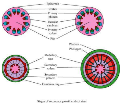
Question 3: Explain the process of secondary growth in stems of woody angiosperm with help of schematic diagrams. What is the significance?
Answer: In woody dicots, the strip of cambium present between the primary xylem and phloem is called the interfascicular cambium. The interfascicular cambium is formed from the cells of the medullary rays adjoining the interfascicular cambium. This results in the formation of a continuous cambium ring. The cambium cuts off new cells toward its either sides. The cells present toward the outside differentiate into the secondary phloem, while the cells cut off toward the pith give rise to the secondary xylem. The amount of the secondary xylem produced is more than that of the secondary phloem. 
The secondary growth in plants increases the girth of plants, increases the amount of water and nutrients to support the growing number of leaves, and also provides support to plants.

Question 4: Draw illustrations to bring out anatomical difference between
(a) Monocot root and dicot root
(b) Monocot stem and dicot stem
Answer: (a) Monocot root and dicot root 

(b) 
Monocot stem and dicot stem


Question 5: Cut a transverse section of young stem of a plant from your school garden and observe it under the microscope. How would you ascertain whether it is a monocot stem or dicot stem? Give reasons.
Answer: The dicot stem is characterised by the presence of conjoint, collateral, and open vascular bundles, with a strip of cambium between the xylem and phloem. The vascular bundles are arranged in the form of a ring, around the centrally-located pith. The ground tissue is differentiated into the collenchyma, parenchyma, endodermis, pericycle, and pith. Medullary rays are present between the vascular bundles.


The monocot stem is characterised by conjoint, collateral, and closed vascular bundles, scattered in the ground tissue containing the parenchyma. Each vascular bundle is surrounded by sclerenchymatous bundle-sheath cells. Phloem parenchyma is absent and water-containing cavities are present.


Question 6: The transverse section of a plant material shows the following anatomical features, (a) the vascular bundles are conjoint, scattered and surrounded by sclerenchymatous bundle sheaths (b) phloem parenchyma is absent. What will you identify it as?
Answer: The monocot stem is characterised by conjoint, collateral, and closed vascular bundles, scattered in the ground tissue containing the parenchyma. Each vascular bundle is surrounded by sclerenchymatous bundle-sheath cells. Phloem parenchyma and medullary rays are absent in monocot stems.

Question 7: Why are xylem and phloem called complex tissues?
Answer: Xylem and phloem are known as complex tissues as they are made up of more than one type of cells. These cells work in a coordinated manner, as a unit, to perform the various functions of the xylem and phloem.
Xylem helps in conducting water and minerals. It also provides mechanical support to plants. It is made up of the following components:
Tracheids (xylem vessels and xylem tracheids)
Xylem parenchyma
Xylem fibres
Tracheids are elongated, thick-walled dead cells with tapering ends. Vessels are long, tubular, and cylindrical structures formed from the vessel members, with each having lignified walls and large central cavities. Both tracheids and vessels lack protoplasm. 
Xylem fibres consist of thick walls with an almost insignificant lumen. They help in providing mechanical support to the plant. Xylem parenchyma is made up of thin-walled parenchymatous cells that help in the storage of food materials and in the radial conduction of water.

Phloem helps in conducting food materials. It is composed of:
Sieve tube elements
Companion cells
Phloem parenchyma
Phloem fibres
Sieve tube elements are tube-like elongated structures associated with companion cells. The end walls of sieve tube elements are perforated to form the sieve plate. Sieve tube elements are living cells containing cytoplasm and nucleus. 
Companion cells are parenchymatous in nature. They help in maintaining the pressure gradient in the sieve tube elements. Phloem parenchyma helps in the storage of food and is made up of long tapering cells, with a dense cytoplasm. Phloem fibres are made up of elongated sclerenchymatous cells with thick cell walls.

Question 8: What is stomatal apparatus? Explain the structure of stomata with a labelled diagram.
Answer: Stomata are small pores present in the epidermis of leaves. They regulate the process of transpiration and gaseous exchange. The stomatal pore is enclosed between two bean-shaped guard cells. The inner walls of guard cells are thick, while the outer walls are thin. The guard cells are surrounded by subsidiary cells. These are the specialised epidermal cells present around the guard cells. The pores, the guard cells, and the subsidiary cells together constitute the stomatal apparatus.


Question 9: Name the three basic tissue systems in the flowering plants. Give the tissue names under each system.
Answer: No.          Tissue system                          Tissues present
1.                             Epidermal tissue system          Epidermis, trichomes, hairs, stomata
2.                             Ground tissue system               Parenchyma, collenchyma, sclerenchyma, mesophyll
3.                            Vascular tissue system              Xylem, phloem, cambium

Question 10: How is the study of plant anatomy useful to us?
Answer: The study of plant anatomy helps us to understand the structural adaptations of plants with respect to diverse environmental conditions. It also helps us to distinguish between monocots, dicots, and gymnosperms. Such a study is linked to plant physiology. Hence, it helps in the improvement of food crops. The study of plant-structure allows us to predict the strength of wood. This is useful in utilising it to its potential. The study of various plant fibres such as jute, flax, etc., helps in their commercial exploitation.

Question 11: What is periderm? How does periderm formation take place in dicot stem?
Answer: Periderm is composed of the phellogen, phellem, and phelloderm.
During secondary growth, the outer epidermal layer and the cortical layer are broken because of the cambium. To replace them, the cells of the cortex turn meristematic, giving rise to cork cambium or phellogen. It is composed of thin-walled, narrow and rectangular cells.
Phellogen cuts off cells on its either side. The cells cut off toward the outside give rise to the phellem or cork. The suberin deposits in its cell wall make it impervious to water. The inner cells give rise to the secondary cortex or phelloderm. The secondary cortex is parenchymatous.

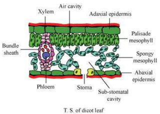
Question 12: Describe the internal structure of a dorsiventral leaf with the help of labelled diagrams.
Answer: Dorsiventral leaves are found in dicots. The vertical section of a dorsiventral leaf contains three distinct parts.
Epidermis: Epidermis is present on both the upper surface (adaxial epidermis) and the lower surface (abaxial epidermis). The epidermis on the outside is covered with a thick cuticle. Abaxial epidermis bears more stomata than the adaxial epidermis.
Mesophyll: Mesophyll is a tissue of the leaf present between the adaxial and abaxial epidermises. It is differentiated into the palisade parenchyma (composed of tall, compactly-placed cells) and the spongy parenchyma (comprising oval or round, loosely-arranged cells with inter cellular spaces). Mesophyll contains the chloroplasts which perform the function of photosynthesis.
Vascular system: The vascular bundles present in leaves are conjoint and closed. They are surrounded by thick layers of bundle-sheath cells.
 (From www.ncrtsolutions.in)
CLASS 11 Biology CHAPTER 16 Digestion and Absorption (NCERT Solution)

CLASS 11 Biology 
CHAPTER 16 
Digestion and Absorption (NCERT Solution)

Question 1: Choose the correct answer among the following:
(a) Gastric juice contains
(i) pepsin, lipase and rennin
(ii) trypsin lipase and rennin
(iii) trypsin, pepsin and lipase
(iv) trypsin, pepsin and renin

(b) Succus entericus is the name given to
(i) a junction between ileum and large intestine
(ii) intestinal juice
(iii) swelling in the gut
(iv) appendix
Answer (a): (i) Pepsin, lipase, and rennin
Gastric juice contains pepsin, lipase, and rennin. Pepsin is secreted in an inactive form as pepsinogen, which is activated by HCl. Pepsin digests proteins into peptones. Lipase breaks down fats into fatty acids. Rennin is a photolytic enzyme present in the gastric juice. It helps in the coagulation of milk.
Answer (b): (ii) Intestinal juice
Succus entericus is another name for intestinal juice. It is secreted by the intestinal gland. Intestinal juice contains a variety of enzymes such as maltase, lipases, nucleosidases, dipeptidases, etc.

Question 2: Match column I with column II
Column I                                              Column II
(a) Bilirubin and biliverdin                   (i) Parotid
(b) Hydrolysis of starch                        (ii) Bile
(c) Digestion of fat                               (iii) Lipases
(d) Salivary gland                                 (iv) Amylases
Answer: Column I                                Column II
(a) Bilirubin and biliverdin                     (ii) Bile
(b)  Hydrolysis of starch                         (iv) Amylases
(c) Digestion of fat                                 (iii) Lipases
(d) Salivary gland                                    (i) Parotid

Question 3: Answer briefly:
(a) Why are villi present in the intestine and not in the stomach?
(b) How does pepsinogen change into its active form?
(c) What are the basic layers of the wall of alimentary canal?
(d) How does bile help in the digestion of fats?
Answer:


 (a) The mucosal wall of the small intestine forms millions of tiny finger-like projections known as villi. These villi increase the surface area for more efficient food absorption. Within these villi, there are numerous blood vessels that absorb the digested products of proteins and carbohydrates,
carrying them to the blood stream. The villi also contain lymph vessels for absorbing the products of fat-digestion. From the blood stream, the absorbed food is finally delivered to each and every cell of the body. The mucosal walls of the stomach form irregular folds known as rugae. These help increase the surface area to volume ratio of the expanding stomach.

(b) Pepsinogen is a precursor of pepsin stored in the stomach walls. It is converted into pepsin by hydrochloric acid. Pepsin is the activated in the form of pepsinogen.

(c) The walls of the alimentary canal are made up of four layers. These are as follows:


(i) Serosa is the outermost layer of the human alimentary canal. It is made up of a thin layer of secretory epithelial cells, with some connective tissues underneath.
(ii) Muscularis is a thin layer of smooth muscles arranged into an outer longitudinal layer and an inner circular layer.
(iii) Sub-mucosa is a layer of loose connective tissues, containing nerves, blood, and lymph vessels. It supports the mucosa.
(iv) Mucosa is the innermost lining of the lumen of the alimentary canal. It is mainly involved in absorption and secretion.

(d) Bile is a digestive juice secreted by the liver and stored in the gall bladder. Bile juice has bile salts such as bilirubin and biliverdin. These break down large fat globules into smaller globules so that the pancreatic enzymes can easily act on them. This process is known as emulsification of fats. Bile juice also makes the medium alkaline and activates lipase.

Question 4: State the role of pancreatic juice in digestion of proteins.
Answer: Pancreatic juice contains a variety of inactive enzymes such as trypsinogen, chymotrypsinogen, and carboxypeptidases. These enzymes play an important role in the digestion of proteins.
Physiology of protein-digestion
The enzyme enterokinase is secreted by the intestinal mucosa. It activates trypsinogen into trypsin.

Trypsin then activates the other enzymes of pancreatic juice such as chymotrypsinogen and carboxypeptidase.
Chymotrypsinogen is a milk-coagulating enzyme that converts proteins into peptides.


Carboxypeptidase acts on the carboxyl end of the peptide chain and helps release the last amino acids. Hence, it helps in the digestion of proteins.


Thus, in short, we can say that the partially-hydrolysed proteins present in the chyme are acted upon by various proteolytic enzymes of the pancreatic juice for their complete digestion.


Question 5: Describe the process of digestion of protein in stomach.
Answer: The digestion of proteins begins in the stomach and is completed in the small intestine. The digestive juice secreted in the gastric glands present on the stomach walls is called gastric juice. The food that enters the stomach becomes acidic on mixing with this gastric juice. The main components of gastric juice are hydrochloric acid, pepsinogen, mucus, and rennin. Hydrochloric acid dissolves the bits of food and creates an acidic medium so that pepsinogen is converted into pepsin. Pepsin is a
protein- digesting enzyme. It is secreted in its inactive form called pepsinogen, which then gets activated by hydrochloric acid. The activated pepsin then converts proteins into proteases and peptides.

Rennin is a proteolytic enzyme, released in an inactive form called prorennin. Rennin plays an important role in the coagulation of milk.


Question 6: Given the dental formula of human beings
Answer: The dental formula expresses the arrangement of teeth in each half of the upper jaw and the lower jaw. The entire formula is multiplied by two to express the total number of teeth.
The dental formula for milk teeth in humans is:
Each half of the upper jaw and the lower jaw has 2 incisors, 1 canine, and 2 molars. Premolars are absent in milk teeth.
The dental formula for permanent teeth in humans is:


Each half of the upper jaw and the lower jaw has 2 incisors, 1 canine, 2 premolars, and 3 molars. An adult human has 32 permanent teeth.

Question 7: Bile juice contains no digestive enzymes, yet it is important for digestion. Why?
Answer: Bile is a digestive juice secreted by the liver. Although it does not contain any digestive enzymes, it plays an important role in the digestion of fats. Bile juice contains bile salts, bile pigments like bilirubin, biliverdin and phospholipids. Bile salts break down large fat globules into smaller globules so that the pancreatic enzymes can easily act on them. This process is known as emulsification of fats. Bile juice also makes the medium alkaline and activates lipase.

Question 8: Describe the digestive role of chymotrypsin. What two other digestive enzymes of the same category are secreted by its source gland?
Answer: The enzyme trypsin (present in the pancreatic juice) activates the inactive enzyme chymotrypsinogen into chymotrypsin.

The activated chymotrypsin plays an important role in the further breakdown of the partially-hydrolysed proteins.


The other digestive enzymes of the same category are trypsinogen and carboxypeptidase. These are secreted by the
same source-gland, pancreas.
Trypsinogen is present in an inactive form in the pancreatic juice. The enzyme enterokinase secreted by the intestinal mucosa – activates trypsinogen into trypsin.

The activated trypsin then further hydrolyses the remaining trypsinogen and activates other pancreatic enzymes such as chymotrypsinogen and carboxypeptidase. Trypsin also helps in breaking down proteins into peptides.


Carboxypeptidases act on the carboxyl end of the peptide chain and help in releasing the last amino acids.

Question 9: 
How are polysaccharides and disaccharides digested?
Answer: The digestion of carbohydrates takes place in the mouth and the small intestine region of the alimentary canal. The enzymes that act on carbohydrates are collectively known as carbohydrases.
Digestion in the mouth:
As food enters the mouth, it gets mixed with saliva. Saliva secreted by the salivary glands contains a digestive enzyme called salivary amylase. This enzyme breaks down starch into sugar at pH 6.8. 

Salivary amylase continues to act in the oesophagus, but its action stops in the stomach as the contents become acidic. Hence, carbohydrate-digestion stops in the stomach.
Digestion in the small intestine:
Carbohydrate-digestion is resumed in the small intestine. Here, the food gets mixed with the pancreatic juice and the intestinal juice. Pancreatic juice contains the pancreatic amylase that hydrolyses the polysaccharides into disaccharides.
Similarly, the intestinal juice contains a variety of enzymes (disaccharidases such as maltase, lactase, sucrase, etc.).
These disaccharidases help in the digestion of disaccharides. The digestion of carbohydrates is completed in the small intestine.

Question 10: What would happen if HCl were not secreted in the stomach?
Answer: Hydrochloric acid is secreted by the glands present on the stomach walls. It dissolves bits of food and creates an acidic medium. The acidic medium allows pepsinogen to be converted into pepsin. Pepsin plays an important role in the digestion of proteins. Therefore, if HCl were not secreted in the stomach, then pepsin would not be activated. This would affect protein digestion. A pH of about 1.8 is necessary for proteins to be digested. This pH is achieved by HCl.

Question 11: How does butter in your food gets digested and absorbed in the body?
Answer: Digestion of fats:
Butter is a fat product and gets digested in the small intestine. The bile juice secreted by the liver contains bile salts that break down large fat globules into smaller globules, so as to increase their surface area for the action of lipase. This process is referred to as emulsification of fats. After this, the pancreatic lipase present in the pancreatic juice and the intestinal lipase present in the intestinal juice
hydrolyse the fat molecules into triglycerides, diglycerides, monoglycerides, and ultimately into glycerol.
Absorption of fats:
Fat absorption is an active process. During fat digestion, fats are hydrolysed into fatty acids and glycerol. However, since these are water insoluble, they cannot be directly absorbed by the blood. Hence, they are first incorporated into small droplets called micelles and then transported into the villi of the intestinal mucosa. They are then reformed into small microscopic particles called chylomicrons, which are small, protein-coated fat globules. These chylomicrons are transported to the lymph vessels in the villi. From the lymph vessels, the absorbed food is finally released into the blood stream and from the blood stream, to each and every cell of the body.

Question 12: Discuss the main steps in the digestion of proteins as the food passes through different parts of the alimentary canal.
Answer: The digestion of proteins begins in the stomach and is completed in the small intestine. The enzymes that act on proteins are known as proteases.
Digestion in the stomach:
The digestive juice secreted in the gastric glands present on the stomach walls is called gastric juice. The main components of gastric juice are HCl, pepsinogen, and rennin. The food that enters the stomach becomes acidic on mixing with this gastric juice. The acidic medium converts inactive pepsinogen into active pepsin. The active pepsin then converts proteins into proteases and peptides.

The enzyme rennin plays an important role in the coagulation of milk.

Digestion in the small intestine:
The food from the stomach is acted upon by three enzymes present in the small intestine, pancreatic juice, intestinal juice (known as succus entericus), and bile juice.
Action of pancreatic juice
Pancreatic juice contains a variety of inactive enzymes such as trypsinogen, chymotrypsinogen, and
carboxypeptidases. The enzymes are present in an inactivated state. The enzyme enterokinase secreted by the intestinal mucosa activates trypsinogen into trypsin.

Action of bile juice
Bile juice has bile salts such as bilirubin and biliverdin which break down large, fat globules into smaller globules so that pancreatic enzymes can easily act on them. This process is known as emulsification of fats. Bile juice also makes the medium alkaline and activates lipase. Lipase then breaks down fats into diglycerides and monoglycerides.

Action of intestinal juice
Intestinal juice contains a variety of enzymes. Pancreatic amylase digests polysaccharides into disaccharides. Disaccharidases such as maltase, lactase, sucrase, etc., further digest the disaccharides.
The proteases hydrolyse peptides into dipeptides and finally into amino acids.

Pancreatic lipase breaks down fats into diglycerides and monoglycerides. The nucleases break down nucleic acids into nucleotides and nucleosides.

Question 13: Explain the term thecodont and diphyodont.
Answer: The codont is a type of dentition in which the teeth are embedded in the deep sockets of the jaw bone. Ankylosis is absent and the roots are cylindrical. Examples include living crocodilians and mammals. Diphyodont is a type of dentition in which two successive sets of teeth are developed during the lifetime of the organism. The first set of teeth is deciduous and the other set is permanent. The deciduous set of teeth is replaced by the permanent adult teeth. This type of dentition can be seen in humans.


Question 14: Name different types of teeth and their number in an adult human.
Answer: There are four different types of teeth in an adult human. They are as follows:
(i) Incisors
The eight teeth in the front are incisors. There are four incisors each in the upper jaw and the lower jaw. They are meant for cutting.
(ii) Canines
The pointy teeth on either side of the incisors are canines. They are four in number, two each placed in the upper jaw and the lower jaw. They are meant for tearing.
(iii) Premolars
They are present next to the canines. They are eight in number, four each placed in the upper jaw and the lower jaw. They are meant for grinding.
(iv) Molars
They are present at the end of the jaw, next to the premolars. There are twelve molars, six each placed in the upper jaw and the lower jaw. Hence, the dental formula in humans is 
This means each half of the upper jaw and the lower jaw has 2 incisors, 1 canine, 2 premolars, and 3 molars. Hence, an adult human has 32 permanent teeth.

Question 15: What are the functions of liver?
Answer: Liver is the largest and heaviest internal organ of the body. It is not directly involved in digestion, but secretes digestive juices. It secretes bile which plays a major role in the emulsification of fats.

(From www.ncrtsolutions.in)

Most Important Ques Asked In Every kind of Exams